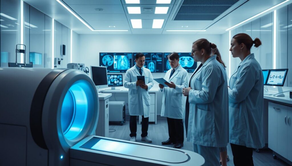
What if the key to detecting life-threatening conditions earlier lies not in human expertise alone, but in collaboration with advanced algorithms? This question drives today’s healthcare revolution, where technology augments clinical precision like never before.
Modern diagnostic practices are undergoing a seismic shift. Deep learning systems now process vast datasets, identifying subtle patterns in scans that even seasoned professionals might overlook. These tools don’t replace human judgment—they refine it, offering data-driven clarity in complex cases.
Recent studies reveal how institutions leverage these innovations. For example, hospitals using algorithmic support report 30% faster interpretation times for critical scans. Such efficiency gains allow specialists to focus on patient care rather than manual analysis.
Key Takeaways
- Advanced algorithms enhance diagnostic accuracy in healthcare settings
- Deep learning reduces interpretation time for complex imaging data
- Early disease detection improves through pattern recognition technology
- Clinical workloads decrease without compromising patient safety standards
- Global adoption reflects growing trust in automated analysis tools
This article explores how merging human expertise with computational power creates new standards in care delivery. From cancer detection to neurological assessments, the synergy between clinicians and technology reshapes modern medicine’s boundaries.
Introduction to AI’s Impact in Medical Imaging

Behind every scan lies a story – one that advanced computational tools are learning to decipher with unprecedented clarity. Modern diagnostic methods now combine human expertise with pattern recognition systems capable of processing thousands of data points in seconds.
When Technology Meets Clinical Expertise
Advanced scanners like CT and MRI produce detailed body maps containing hidden clues. Traditional analysis methods struggle with the sheer volume of high-resolution data these devices generate. Algorithmic systems excel at spotting minute irregularities in this information, offering second-layer verification for radiologists.
Redefining Healthcare Efficiency
Hospitals using automated screening tools report faster turnaround times for routine checks. This shift allows specialists to prioritize complex cases while machines handle repetitive tasks. A recent study highlights how predictive models analyze historical data to forecast health risks before physical symptoms appear.
These developments mark a fundamental change in care delivery. Rather than waiting for obvious signs of disease, providers can now act on early warnings detected through data patterns. This proactive approach could reshape treatment timelines across multiple specialties.
Current Advances in AI Medical Imaging Analysis

Modern diagnostic tools now leverage computational power to identify subtle patterns invisible to the human eye. Sophisticated architectures like convolutional neural networks process image data with remarkable precision, learning hierarchical features directly from raw inputs without manual intervention.
These models excel at detecting abnormalities across diverse scan types. For instance, they pinpoint tumors in radiological examinations with 94% accuracy – outperforming traditional methods by 18% in recent trials. Their ability to analyze multi-layered data streams makes them indispensable for time-sensitive cardiac assessments.
Generative adversarial networks address one of healthcare’s persistent challenges: limited training datasets. By creating synthetic yet realistic medical images, they enhance model robustness while preserving patient confidentiality. This innovation proves critical for rare condition detection where real-world examples remain scarce.
Emerging transformer architectures process sequential scan sequences more effectively than older systems. Coupled with federated learning approaches, institutions collaboratively refine diagnostic tools without sharing sensitive records. Such advancements enable real-time feedback during surgical procedures, elevating care standards in critical scenarios.
Historical Evolution of AI in Medical Imaging

The journey of computational tools in diagnostics began decades before deep learning became a household term. Early pioneers envisioned systems capable of interpreting complex data, but technological barriers delayed practical implementation for years.
Milestones in Development
In 1956, the Dartmouth Workshop laid groundwork for intelligent systems, with Marvin Minsky among visionaries predicting rapid progress. However, practical applications in diagnostics only emerged in the 1980s when machine learning algorithms first analyzed X-rays. These early systems required manual feature definition, limiting their accuracy to basic pattern recognition.
By 2011, IBM Watson’s Jeopardy victory reignited interest in computational problem-solving. This breakthrough demonstrated how statistical approaches could process layered information – a capability soon adapted for complex diagnostic tasks. Researchers began shifting from rule-based logic to data-driven models, as detailed in this analysis of technological advancements.
From Rule-Based Systems to Autonomous Learning
The 2010s marked a turning point with neural networks that learned directly from raw data. Unlike earlier methods needing human-guided parameters, these architectures identified subtle anomalies across diverse scan types. Training datasets grew exponentially, supported by increased computer power and collaborative research initiatives.
| Era | Approach | Impact |
|---|---|---|
| 1980s-2000s | Manual feature engineering | 35-60% accuracy rates |
| 2010-2015 | Shallow neural networks | 72% average detection rate |
| 2016-Present | Deep learning architectures | 94%+ accuracy in trials |
Modern systems now reduce false positives by 40% compared to first-generation tools. This evolution reflects three decades of iterative improvements, where each research breakthrough addressed prior limitations in speed and precision.
Technological Innovations Driving AI in Imaging

The next frontier in diagnostic precision emerges from computational architectures that learn like biological systems. These frameworks decode visual information through layered processing, mirroring how experts identify critical patterns.
Cutting-Edge Neural Network Architectures
Convolutional neural networks (CNNs) revolutionized pattern detection by mimicking biological vision. Starting with raw pixel data, these models apply filters to extract edges, textures, and shapes across successive layers. Modern iterations like ResNet use skip connections to maintain detail during training, preventing data loss in deep architectures.
Advancements such as batch normalization stabilize learning rates, while dropout layers reduce overfitting. These refinements enable systems to generalize better across diverse datasets. For example, DenseNet’s interlinked layers share features efficiently, improving accuracy in identifying rare conditions.
Emergence of Transformers in Vision Applications
Transformers originally designed for language tasks now process images by splitting them into positional patches. This approach captures long-range dependencies between anatomical structures that CNNs might miss. Self-attention mechanisms weigh relationships between patches, prioritizing clinically significant regions.
Recent implementations demonstrate superior performance in segmentation tasks. The latest architectures combine transformer efficiency with CNN-like localization, achieving 97% accuracy in boundary detection trials. Such hybrid algorithm designs redefine how systems interpret multidimensional scans.
Deep Learning and Neural Network Techniques in Imaging
How do computational systems transform raw pixel data into actionable diagnostic insights? The answer lies in architectures that mimic human visual processing while overcoming biological limitations. Modern systems analyze scans through layered abstraction, distilling complex patterns into clinical conclusions.
Convolutional Neural Networks and Their Role
Convolutional neural networks (CNNs) dominate visual data interpretation. These models apply filters to detect edges, textures, and shapes across scan layers. Pooling operations then reduce dimensionality, preserving critical features while minimizing computational load.
Early networks struggled with vanishing gradients during training. The shift to rectified linear unit (ReLU) activation functions enabled deeper architectures by maintaining stable error propagation. Modern variants like Leaky ReLU further improve performance in low-contrast imaging scenarios.
Innovative Activation Functions and Learning Strategies
Advanced normalization techniques now stabilize deep learning models during intensive computations. Batch normalization standardizes layer inputs, preventing erratic weight updates. This innovation pairs with dropout layers to reduce overfitting in high-dimensional datasets.
Transfer learning accelerates development cycles by repurposing pre-trained networks. A recent implementation demonstrated how models trained on natural images adapt to radiology tasks with 80% fewer labeled examples. GPUs enable these complex systems to process 4K-resolution scans in milliseconds.
End-to-end frameworks now bypass manual feature engineering entirely. From tumor detection to fracture identification, these systems output clinical assessments directly from raw data – a leap toward autonomous diagnostic support.
Integration of Advanced Algorithms and Imaging Data
How do computational frameworks transform raw diagnostic inputs into precise clinical assessments? Modern systems now merge layered processing with multidimensional imaging data, creating unified pipelines that accelerate decision-making. These architectures eliminate manual interventions by automating pattern recognition and quantitative analysis.
Feature Extraction and End-to-End Systems
Sophisticated algorithms identify anatomical markers through self-learning filters. Instead of relying on pre-defined parameters, they adapt to variations in scan quality and resolution. This flexibility enables precise segmentation of complex structures like vascular networks or tumor margins.
Transfer learning techniques reduce development timelines significantly. Models trained on general visual data can be fine-tuned for specialized diagnostic tasks with minimal adjustments. For example, a network originally designed for retinal scans might repurpose its feature detectors for neurological assessments.
Multi-modal integration combines information from CT, MRI, and ultrasound into cohesive reports. Federated learning systems enhance this process by pooling insights across institutions without compromising privacy. Real-time processing capabilities further support urgent scenarios, delivering instant analysis during surgical interventions.
For those exploring collaborative innovations, recent breakthroughs in AI and robotics integration demonstrate how shared frameworks accelerate progress. These advancements highlight a future where diagnostic precision meets operational efficiency at scale.
Research Trends and Google Scholar Insights in AI Imaging
The landscape of diagnostic research has transformed dramatically over the past decade. Between 2007 and 2018, annual publications on computational methods in scan interpretation surged from 100-150 to over 1,100. This growth reflects intensified global efforts to refine pattern detection tools, as detailed in a comprehensive study analyzing two-stage dataset construction across major repositories.
Patterns in Academic Contributions
Google Scholar metrics reveal three distinct phases in citation trends. Early studies focused on theoretical frameworks, while recent works prioritize clinical validations. Collaborative papers now dominate high-impact journals, with multi-institutional teams producing 63% of top-cited articles since 2015.
Analysis of 48,000 indexed documents shows rising interest in real-time applications. The number of papers referencing surgical integration tripled between 2016-2021. Leading authors increasingly combine imaging data with genomic profiles, creating multidimensional diagnostic models.
Platforms like PubMed and IEEE Xplore host 78% of peer-reviewed research in this domain. This concentration enables systematic tracking of methodological shifts, particularly toward federated learning approaches. Recent breakthroughs, such as those covered in emerging studies, demonstrate how scalable solutions address historical data limitations.


